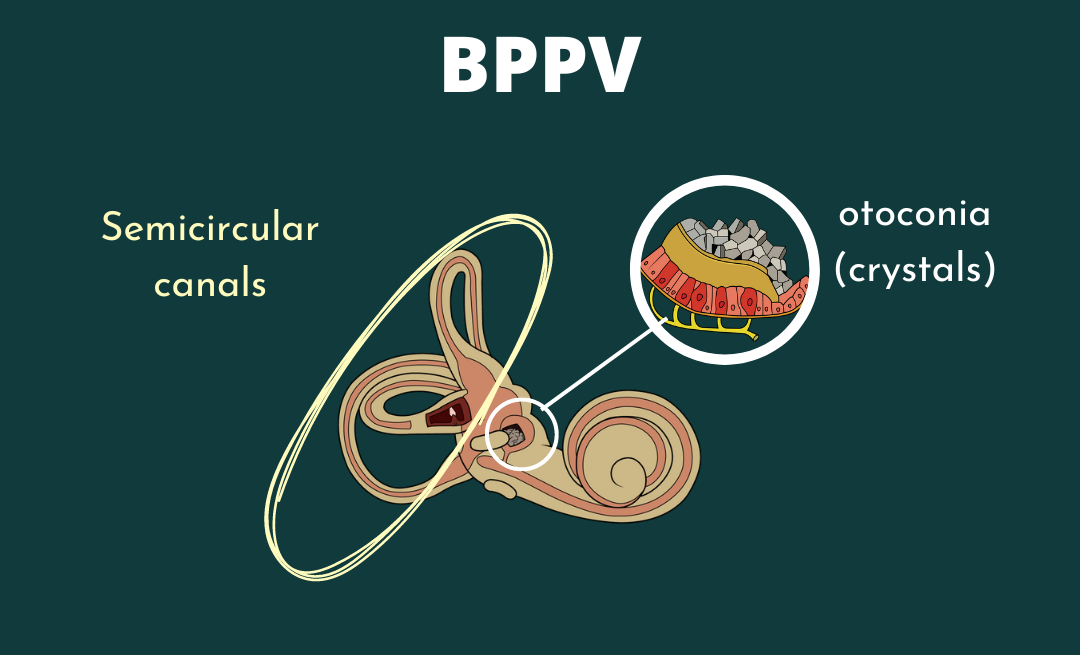
Diagnosing Benign Paroxysmal Positional Vertigo

BPPV, Benign Paroxysmal Positional Vertigo, can be easy to diagnose subjectively, but watching eyes can be difficult. The following is a chart illustrating the direction of eye movement, position of your patient’s head, and the corresponding diagnosis.
As a provider, you need to know what you’re looking at so you know what you’re looking for! In this chart, you will see which type of nystagmus corresponds with which position and which specific BPPV diagnosis.
Please know, when looking at this chart, that it will help you determine the proper diagnosis based on the position and nystagmus. However, if the position and nystagmus don’t seem to match, there is not a crescendo up and down of nystagmus, and a delay does NOT exist upon lying down… it is likely NOT BPPV!
| BPPV Diagnosis | Nystagmus | Position |
| Right Posterior Canal Canalithiasis | Right Upbeating Torsional Nystagmus | Right Dix-Hallpike Position |
| Left Posterior Canal Canalithiasis | Left Upbeating Torsional Nystagmus | Left Dix-Hallpike Position |
| Right Horizontal Canal Canalithiasis | More intense Right Geotropic Nystagmus | Right Roll Test (compared to Left Roll Test Position) |
| Left Horizontal Canal Canalithiasis | More intense Left Geotropic Nystagmus | Left Roll Test (compared to Right Roll Test position) |
| Right Horizontal Canal Cupulolithiasis | Less intense Right Ageotropic & more intense Left Ageotropic nystagmus | Right or Left Roll Test position respectively |
| Left Horizontal Canal Cupulolithiasis | Less intense Left Ageotropic & more intense Right Ageotropic nystagmus | Left or Right Roll Test position Respectively |
| Right Anterior Canal Canalithiasis | Downbeating (and sometimes torsional) Nystagmus | Left Dix-Hallpike Position |
| Left Anterior Canal Canalithiasis | Downbeating (and sometimes torsional) Nystagmus | Right Dix-Hallpike Position |